
Case Gallery
-
CASE 1:
Treatment of a lower incisor showing two canals that join into one canal in the apical one third -
CASE 2:
Treatment of a symptomatic lower molar with an unobturated distal canal and a screw post present -
CASE 3:
Treatment of a failing upper central incisor with a labial sinus tract and radiographic evidence of periradicular inflammatory resorption -
CASE 4:
Treatment of a lower first molar -
CASE 5:
Treatment of an upper lateral incisor -
CASE 6:
Implants -
CASE 7:
Bone Graft
CASE 1
Treatment of a lower incisor showing two canals that join into one canal in the apical one third
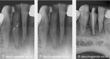 Lower incisors may suffer from post endodontic treatment disease. Approximatly 40% of lower incisors have two canals! The above off angle radiograph demonstrates this second canal. Clinicians often only identifiy a single canal (normally the facial/labial canal) and subseqently leave behind a uncleaned second canal. Leaving unexplored root canal systems leaves the potential to cause post tretment disease and ultimatly a failing endodontic case.
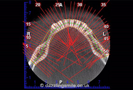
 Here at St Pauls we routinly use low dose sectional Cone Beam CT imaging in pre endodontic assessment which can show these complexities within root canal systems. With the aid of an operating Microscope giving us unsupassed vision deep into the pulp chamber, we are able to explore and fill these hidden systems.
Below is a pre operative CBCT section showing a lower lateral incisor mid root. This demonstrates the ability of CBCT to give us an insight in to root canal anatomy so we can fully identify all the root canal systems.
![]()
CASE 2
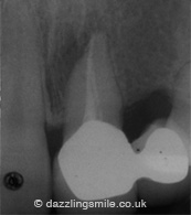
Treatment of a symptomatic lower molar with an unobturated distal canal and a screw post present
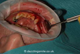
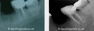 The endodontic treatment options for this case are either non surgical re-root canal treatment (NSRCT) or surgical root canal treatment. NSRCT should always be attempted if it is feasible. However, if it is deemed that the disassembly of the existing restoration is too complex and may lead to damage to the remaining tooth then surgical endodontics maybe preferred.
In this case NSRCT was feasible. I used ultrasonics to remove the Dentatus post contained in the distal canal. The mesial canal shows only one canal to be filled and this filled canal was short of the radiographic apex.
The post treatment radiograph shows the missed mesial canal to be fully obturated and the short mesial canal re treated and obturated further towards the radiographic apex. The distal canal was cleaned, shaped and obturated.
![]()
CASE 3
Treatment of a failing upper central incisor with a labial sinus tract and radiographic evidence of periradicular inflammatory resorption
The pre-operative radiograph shows voids present within the obturated root canal. A periradicular radiolucency with apical resorption is present.
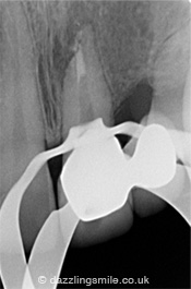
The working radiograph shows an apical plug of MTA in place. The apical constriction had been destroyed due to the resorption. The canal could not be obturated with the conventional methods of gutta purcha
The post-operative radiograph shows a fully obturated root canal system. Clinically the sinus tract disappeared.
![]()
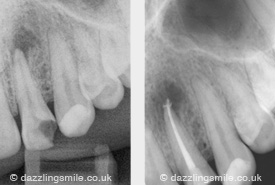
CASE 4
Treatment of a lower first molar
The pre-operative radiograph shows a GP point tracking the sinus to a large periradicular lesion.
The post-operative radiograph shows four fully obturated canals. The sinus tract had healed prior to the obturation of the tooth.
This lower first molar demonstrates how curved canals can be easily shaped, clean and obturated using NiTi rotary files.
![]()
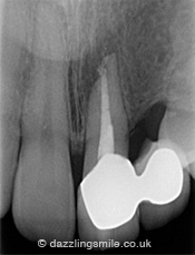
CASE 5
Treatment of an upper lateral incisor
 The post-operative radiograph demonstrating multiple apical portals that were effectively obturated using three dimensional thermal obturation techniques.
![]()
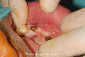
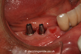
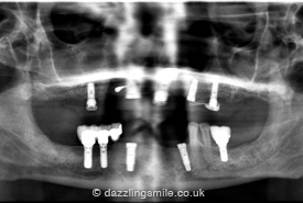
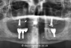
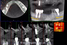
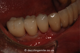
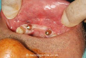
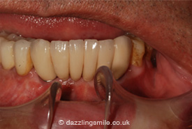
CASE 6
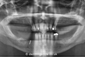
Implants
 |
 |
 |
 |
 |
 |
 |
 |
![]()
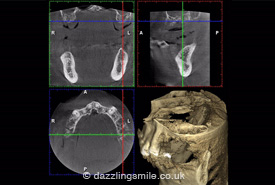
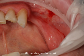
CASE 7
Bone Graft
 |
 |
 |
 |
 |
|
![]()